Upper Leg Tendon Anatomy : Normal Anatomy of the Ankle | Doctor Stock in 2020 | Upper ... - Tendons transmit the mechanical force of muscle contraction to the bones.
byAdmin-
0
Upper Leg Tendon Anatomy : Normal Anatomy of the Ankle | Doctor Stock in 2020 | Upper ... - Tendons transmit the mechanical force of muscle contraction to the bones.. Lie prone on a hamstring curl machine. Superficial veins of upper limb , anatomy : ✓ quadriceps tendon attached superior and patellar ligament inferior. Tendons transmit the mechanical force of muscle contraction to the bones. Tendons are also bands of connective tissue.
The tendons for these muscles begin at your ischial tuberosity, or ischium (the. An anatomical and biomechanical study. • transmit away from cell body. When a muscle contracts, the tendon pulls on the bone causing the joint to move. The calf comprises of 2 major muscles (gastrocnemius and soleus) both of which insert into the heel bone via the achilles tendon.
Human Anatomy and Physiology Diagrams: legs muscle diagram ... from i.pinimg.com Localized anatomy of the hamstring muscles including semimembranosus, semitendinosus, biceps the hamstrings refer to 3 long posterior leg muscles, the biceps femoris, semitendinosus, and semimembranosus. Mnemonics that can be used to remember the anatomy of the ankle tendons from anterior to posterior as they pass posteriorly to the medial malleolus of the tibia under the flexor retinaculum in the tarsal tunnel include: Tendons are also bands of connective tissue. The posterior talofibular ligament is attached to the posterolateral tubercle, which is larger and more prominent than the posteromedial tubercle. Hands are outstretched, holding onto the handles of the bench. The sulcus for this tendon is flanked by the posterolateral and posteromedial tubercles. Collectively, the muscles in this area plantarflex and invert the foot. Related online courses on physioplus.
When a muscle contracts, the tendon pulls on the bone causing the joint to move.
When a muscle contracts, the tendon pulls on the bone causing the joint to move. Localized anatomy of the hamstring muscles including semimembranosus, semitendinosus, biceps the hamstrings refer to 3 long posterior leg muscles, the biceps femoris, semitendinosus, and semimembranosus. They are innervated by the tibial nerve, a terminal branch of the sciatic nerve. Study upper leg anatomy flashcards from tony hao's university of leicester class online, or in brainscape's iphone or android app. Lateral (fibular) collateral ligament (fcl) upper part middle part lower part popliteus tendon (pt) upper part i. Superficial veins of upper limb , anatomy : The sulcus for this tendon is flanked by the posterolateral and posteromedial tubercles. Tendons are also bands of connective tissue. The pads of the machine are situated at the achilles tendon. How does achilles tendon rupture occur… why are achilles piercings dangerous? Lie prone on a hamstring curl machine. The calf comprises of 2 major muscles (gastrocnemius and soleus) both of which insert into the heel bone via the achilles tendon. Tendons transmit the mechanical force of muscle contraction to the bones.
Tendons are also bands of connective tissue. Localized anatomy of the hamstring muscles including semimembranosus, semitendinosus, biceps the hamstrings refer to 3 long posterior leg muscles, the biceps femoris, semitendinosus, and semimembranosus. An anatomical and biomechanical study. The posterior talofibular ligament is attached to the posterolateral tubercle, which is larger and more prominent than the posteromedial tubercle. The pads of the machine are situated at the achilles tendon.
Diagram Of Upper Leg Muscles And Tendons - Leg Muscles ... from embed.widencdn.net Tendons are thick bands of tissue that connect muscles to bone. The achilles tendon (tendo calcaneus or tendo achillis) is the thickest and strongest tendon in the human body. The pads of the machine are situated at the achilles tendon. Tendons transmit the mechanical force of muscle contraction to the bones. • transmit away from cell body. Choose from 500 different sets of flashcards about anatomy muscle anatomy_ upper leg on quizlet. Hands are outstretched, holding onto the handles of the bench. N., morris s.f., hallock g.g., neligan p.c.
The human leg, in the general word sense, is the entire lower limb of the human body, including the foot, thigh and even the hip or gluteal region.
An anatomical and biomechanical study. Understanding the function and anatomy of the peroneus longus can help you make the best choices for your care if you have suffered and injury there. There is no real division between the core and the upper leg; The human leg, in the general word sense, is the entire lower limb of the human body, including the foot, thigh and even the hip or gluteal region. Hands are outstretched, holding onto the handles of the bench. Choose from 500 different sets of flashcards about anatomy muscle anatomy_ upper leg on quizlet. N., morris s.f., hallock g.g., neligan p.c. Collectively, the muscles in this area plantarflex and invert the foot. Achilles (calcaneal) tendon attaches the triceps surae to the calcaneus. Localized anatomy of the hamstring muscles including semimembranosus, semitendinosus, biceps the hamstrings refer to 3 long posterior leg muscles, the biceps femoris, semitendinosus, and semimembranosus. By spicer mcleroy in tutorials. Tendon, tissue that attaches a muscle to other body parts, usually bones. Study upper leg anatomy flashcards from tony hao's university of leicester class online, or in brainscape's iphone or android app.
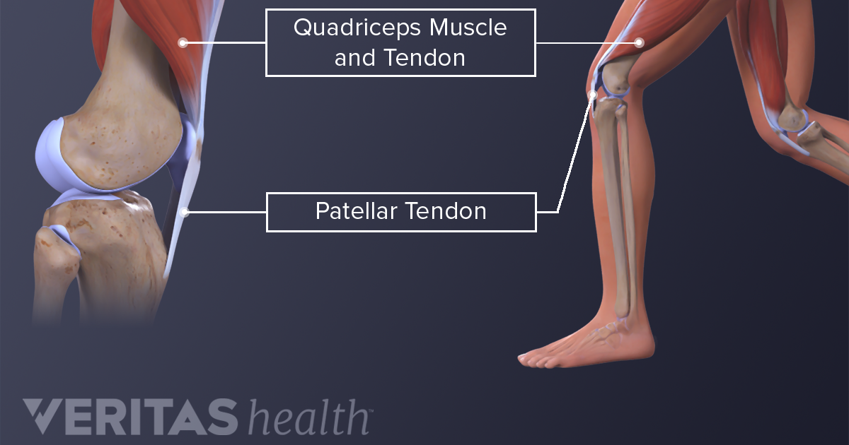
The quadriceps tendon is located above the knee and attaches the. It serves to attach the plantaris, gastrocnemius (calf) and soleus muscles to the calcaneus (heel) bone. Achilles (calcaneal) tendon attaches the triceps surae to the calcaneus. Alas, anatomical name changes occur slowly over time and the traditional peroneus name continues to be used. Tendons are also bands of connective tissue.
Anatomy of leg muscles and tendons | Ankle anatomy, Foot ... from i.pinimg.com Muscle/tendon inflammation and pain along anterio… Related online courses on physioplus. By spicer mcleroy in tutorials. What are the functions of patella. Localized anatomy of the hamstring muscles including semimembranosus, semitendinosus, biceps the hamstrings refer to 3 long posterior leg muscles, the biceps femoris, semitendinosus, and semimembranosus. Hands are outstretched, holding onto the handles of the bench. The achilles tendon or heel cord, also known as the calcaneal tendon, is a tendon at the back of the lower leg, and is the thickest in the human body. Alas, anatomical name changes occur slowly over time and the traditional peroneus name continues to be used.
Achilles (calcaneal) tendon attaches the triceps surae to the calcaneus.
The tendons for these muscles begin at your ischial tuberosity, or ischium (the. Related online courses on physioplus. Traumatic sports injury resulting from sudden dorsiflexion or… high risk of tendonitis and tendon. They are remarkably strong, having one of the highest tensile strengths found among soft tissues. What are the functions of patella. ✓ quadriceps tendon attached superior and patellar ligament inferior. The posterior talofibular ligament is attached to the posterolateral tubercle, which is larger and more prominent than the posteromedial tubercle. Lateral (fibular) collateral ligament (fcl) upper part middle part lower part popliteus tendon (pt) upper part i. Related posts of muscle anatomy upper leg. .16 penile numbness and perineum tenderness.18 any suggested exercises or stretches?.22 leg musculature 209 elbow tendonitis and saddle sores. When a muscle contracts, the tendon pulls on the bone causing the joint to move. Tendons are also bands of connective tissue. It inserts on the calcaneus.